|
|
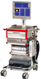
Information box |
The main purpose of this site is to extend the
intraoperative monitoring to include the neurophysiologic
parameters with intraoperative navigation guided with Skyra 3
tesla MRI and other radiologic facilities to merge the
morphologic and histochemical data in concordance with the
functional data.
 CNS Clinic
Located in Jordan Amman near Al-Shmaisani hospital, where all
ambulatory activity is going on.
Contact: Tel: +96265677695, +96265677694.
Skyra running
A magnetom Skyra 3 tesla MRI with all clinical applications
started to run in our hospital in 28-October-2013.
Shmaisani hospital
The hospital where the project is located and running diagnostic
and surgical activity. |
|
 |
|
|
 |
Tim
Planning Suites |
 |
Easy planning of extended Field of View examinations
in an efficient way using Set-n-Go protocols. It allows
planning of several stations at once e.g. on composed
localizer images. The overlap of slice groups can be
adjusted. All stations can have independent parameter
settings although they are displayed together. A special
coupling mode allows easy positioning of all stations at
once according to the patient’s anatomy. Fully supports
scan@center and Phoenix functionality.
• Tim Planning UI with optimized layout for slice
positioning
• Ready to use Set-n-Go protocols for different clinical
questions
• Integrated toolbar for fast advanced slice planning:
FoV-Plus, FoV-Minus, AlignParallel, AlignFieldOfViews.
|
|
Tim
Application Suites |
|
The Tim Application Suite offers a complete range of
clinically optimized sequences, protocols and workflow
functionalities for all body regions. Excellent
head-to-toe imaging can be accomplished with the
sequences and features included in this application
suite. To enable this comprehensive application range,
nine dedicated application packages have been included.
Neuro Suite
Angio Suite
Cardiac Suite
Body Suite
Whole
Body Suite
Onco Suite
Breast Suite
Ortho Suite
Pediatric Suite
Scientific Suite
 |
Neuro Suite |
 |
Comprehensive head and
spine examinations can be performed with
dedicated programs. High resolution protocols
and fast protocols for uncooperative patients
are provided. The Neuro Suite also includes
protocols for diffusion imaging, perfusion
imaging, and fMRI. It includes for example:
- EPI sequences and protocols for
diffusion, perfusion and fMRI for advanced
neurological applications.
Diffusion weighted imaging is possible with up
to 16 b-values in the orthogonal directions.
Dynamic Analysis software (included in standard
configuration) enables calculation of:
- ADC maps
- t-test maps from the EPI images for fMRI
- Time-to-Peak maps for perfusion
analysis.
- Whole spine protocols acquire in
multiple steps via software controlled table
movement in a single click.
- 3D isotropic resolution volume imaging
using T1 3D MPRAGE / 3D FLASH, SPACE DarkFluid,
T2 SPACE and 3D TSE
- T2-weighted high resolution 3D Restore
protocols optimized for inner ear examinations
- Whole-spine protocols in multiple steps
with software controlled table movement
- 2D and 3D MEDIC protocols for
T2-weighted imaging, particularly for C-spine
examinations in axial orientation where
reproducibility is difficult due to CSF
pulsations and blood flow artifacts
- 3D Myelograms with 3D HASTE and 3D
True-FISP for anatomical details
- Dynamic sacro-iliac joint imaging after
contrast administration using a fast T1-weighted
FLASH 2D sequence
- Spine diffusion protocols to
differentiate osteoporosis versus tumor
infiltration and post-radiotherapy changes
versus residual tumor with PSIF sequence
- Precision filter for high spatial
accuracy e.g. for neuro intra-operative imaging
and stereotactic planning
- 3D CISS (Constructive Interference in
Steady State) for excellent visualization of
fine structures such as cranial nerves. High
resolution imaging of inner ear and spine
- AutoAlign Head LS providing a fast,
easy, standardized, and reproducible patient
scanning supporting reading by delivering a
higher and more standardized image quality.
|
|
Angio Suite |
|
Excellent MR
Angiography can be performed to visualize
arteries and veins with or without contrast
agent.
Contrast-enhanced MRA
- 3D contrast-enhanced MRA protocols for
e.g. single step, dynamic, peripheral, whole
body MRA with the shortest TR and TE. The strong
gradients make it possible to separate the
arterial phase from the venous phase.
- TestBolus workflow for optimized bolus
timing and superb image quality.
- CareBolus functionality for accurate
determination of the bolus arrival time and the
"Stop and Continue" of the 3D ce-MRA protocol
after the 2D bolus control scan.
- Dynamic ce-MRA for 3D imaging over time.
Non-contrast-MRA and venography
- 2D and 3D Time-of-Flight (ToF) protocols
for MRA for the Circle of Willis, carotids, neck
vessels, and breath-hold protocols for abdominal
vessels
- Triggered 2D ToF sequences for
non-contrast MRA, particularly of the abdomen
and the extremities
- 2D/3D Phase-Contrast
- MR venography with 2D/3D Time-of-Flight
(ToF) and Phase-Contrast
- TONE (Tilted Optimized Non-saturation
Excitation) and MTC (Magnetization Transfer
Contrast) techniques for improved
Contrast-to-Noise Ratio (CNR)
 Image processing tools
- MPR, MIP, MinIP, and 3D SSD
- Inline MIP for immediate results
- Inline subtraction of pre- and
post-contrast measurements
- Inline standard deviation maps of
Phase-Contrast measurements for delineation of
arteries and veins
|
|
Cardiac Suite |
|
The cardiac suite covers
comprehensive 2D routine cardiac applications,
ranging from morphology and ventricular function
to tissue characterization. Featuring syngo BEAT
2D in conjunction with iPAT and T-PAT
techniques.
Cardiac views
- Fast acquisition of the basic cardiac
orientations for further examination planning
- Cardiac scouting provides users with a
step-by-step procedure for the visualization and
planning of typical cardiac views, e.g. based on
TrueFISP or Dark Blood TurboFLASH: short axis,
4-chamber and 2-chamber views.
syngo BEAT
- Unique tool for fast and easy
cardiovascular MR imaging
- E.g. 1 click change from FLASH to
TrueFISP for easy contrast optimization
- 1-click to switch arrhythmia rejection
on / off
- 1-click change from Cartesian to radial
sampling to increase effective image resolution
(e.g. in pediatric patients) and avoid folding
artifacts in large patients
Visualization of structural cardiovascular
pathologies with CMR – syngo BEAT
- Breath-hold and free breathing
techniques for strong contrast between the blood
and vascular structures. Dark Blood TSE and
HASTE imaging are available for the structural
evaluation of the cardiothoracic anatomy,
including vessels or heart valves. Cine
techniques (FLASH & TrueFISP) for
high-resolution valve evaluation
- Multiple contrasts such as T1- and
T2-weighted imaging for use in diseases such as
myocarditis (inflammation / hyperaemia), ARVD
(fibrous-fatty degeneration) or acute myocardial
infarction (edema)
- Dark-blood TSE with motion compensation
for high-quality vessel wall imaging in small or
large vessels
Tools for rapid evaluation of left or right
ventricular function
- Acquisition of a stack of short-axis
slices (standard segmented FLASH, or advanced
segmented TrueFISP)
- Automatic adjustment of the acquisition
window to the current heart rate
- Use of the Inline ECG for graphical ECG
triggering setup
- Retrospective gating with cine sequences
(TrueFISP, FLASH)
- Protocols for whole-heart coverage
- iPAT integration for highest temporal
and spatial resolution
- Real-time imaging in case the patient is
not able to hold his breath
Dynamic imaging and tissue characterization with
syngo BEAT
- Protocols for high-contrast and
high-resolution tissue characterization
- Protocols for stress and rest imaging
with TrueFISP or TurboFLASH contrast support the
acquisition of multiple slices with high
resolution and arbitrarily adjustable slice
orientation for each slice
- T-PAT with mSENSE and GRAPPA for
advanced parallel imaging provides fast
high-resolution dynamic imaging
- Segmented IR TrueFISP / FLASH with TI
scout for optimization of tissue contrast
- Advanced tissue characterization with 2D
phase-sensitive IR (PSIR) sequences TrueFISP and
FLASH contrast. Magnitude and phase-sensitive
images with one acquisition
- Simple: no adjustment of inversion time
(TI) necessary with PSIR technique
- Ungated single-shot PSIR imaging for
tissue characterization under difficult
conditions: free-breathing technique that can be
applied even in case of arrhythmia
Physiological Measurement Unit (PMU) - Wireless
Physio Control
- Synchronizes the measurement with the
physiological cycles (triggering to minimize
motion artifacts caused by cardiac and
respiratory movements)
- Wireless Sensors
- Wireless Vector ECG / respiration and
pulse sensors for physiologically synchronized
imaging, rechargeable battery-powered - for
optimized patient handling
- Physiological Signals Display
- ECG (3 channels)
- Pulse
- Respiration
- External Trigger Input Display
ECG Triggering:
- Acquisition of multiple slices, e.g. of
the heart, at different phases of the cardiac
cycle
- Excellent image quality by synchronizing
data acquisition with cardiac motion
- Peripheral Pulse Triggering:
- Reduces flow artifacts caused by
pulsatile blood flow
- Excellent image quality by synchronizing
data acquisition to the pulsatile blood flow
- Respiratory Triggering:
- Excellent image quality by synchronizing
data acquisition with the respiratory motion
- External Triggering:
- Interface for trigger input from
external sources (e.g. Patient Monitoring
System) inside the examination room
- Interface for trigger input from
external sources (e.g. pulse generator, trigger
sources for fMRI) outside the examination room
- Optical trigger output for fMRI
- Retrospective gating for ECG, peripheral
pulse, and external trigger input
|
|
Breast Suite |
|
MR imaging has proven a
very high sensitivity for breast lesions and is
the gold standard for the examination of
silicone implants. Extremely high spatial and
temporal resolution can be achieved in very
short measuring times by using iPAT with GRAPPA.
Excellent soft tissue differentiation,
customized protocols (e.g. with fat saturation
or water excitation or silicone excitation), as
well as flexible multiplanar visualization allow
for fast, simple and reproducible evaluation of
MR breast examinations.
This package includes:
- Quantitative evaluation and fast
analysis of the data with colorized Wash-in,
Wash-out, Time-To-Peak,
Positive-Enhancement-Integral, MIPtime and
combination maps with Inline technology or for
offline calculation
- High-resolution 2D protocols for
morphology evaluation
- High-resolution 3D protocols covering
both breasts simultaneously
- Protocols to support interventions (fine
needle and vacuum biopsies, wire localization)
- Protocols for evaluating breasts with
silicone implants
- Automatic and manual frequency
adjustment, taking into account the silicone
signal
- Detection of the silicone signal either
to suppress the silicone signal, if the
surrounding tissue is to be evaluated, or to
suppress the tissue signal in order to detect an
implant leakage
- SPAIR - robust fat sat (robust fat
suppression using an adiabatic frequency
selective inversion pulse)
- DIXON - 2-point Dixon with 3D VIBE, the
following contrasts can be obtained: in-phase,
opposed phase, fat and water image.
- iPAT with GRAPPA for maximum resolution
in short time
- Inline subtraction and MIP display
- Offline subtraction, MPR and MIP display
- syngo REVEAL: diffusion imaging for
breast exams
- iPAT Extension that allows
state-of-the-art sagittal breast imaging
- iPAT Extension allows bilateral 3D
sagittal breast imaging with Fat Sat or Water
excitation
The Breast Suite also includes:
syngo VIEWS (Volume Imaging with Enhanced Water
Signal)
- bilateral - both breasts are examined
simultaneously
- axial - the milk ducts are directly
displayed
- fat-saturated or water-excited - fat
complicates clinical evaluation and is
suppressed
- near-isotropic 3D measurement - the same
voxel size in all three directions for
reconstruction in any slice direction
- submillimeter voxel - highest resolution
for precise evaluation
|
|
Body Suite |
|
Body Suite covers your
needs for clinical body applications. Ultrafast
high resolution 2D and 3D protocols are provided
for abdomen, pelvis, MR Colonography, MRCP,
dynamic kidney, and MR Urography applications.
Siemens unique 2D PACE technique makes body
imaging easy allowing for multi-breath hold
examinations as well as free breathing during
the scans. Motion artifacts are greatly reduced
with 2D PACE Inline technology.
This package includes:
- Free breathing 2D PACE applications with
2D/3D HASTE (RESTORE) and 2D/3D TSE (RESTORE)
- Optimized fast single shot HASTE
protocols and high-resolution 3D RESTORE
protocols based on SPACE and TSE for MRCP and MR
Urography examinations
ABDOMEN:
2D:
- T1w (FLASH) breath-hold scans +/- Fat
Sat (SPAIR, Q-FatSat, in-/opp-phase)
- T2w (HASTE, TSE/BLADE, EPI ) breath-hold
scans +/- Fat Sat (SPAIR, FatSat, STIR)
- T1w (TFL) triggered scans (2D PACE free
breathing) in-/opp-phase
- T2w (HASTE, TSE/BLADE, EPI)
triggered scans (2D PACE free breathing) +/- Fat
Sat (SPAIR,FatSat, STIR) as well as HASTE- and
TSE-multi-echo
- Optimized fast single shot HASTE
protocols and high-resolution 3D RESTORE
protocols based on SPACE and TSE for MRCP and MR
urography examinations
3D:
- Dixon (VIBE 2pt-Dixon) breath-hold
scans, following contrasts can be obtained:
in-phase, opposed phase, fat and water image.
- Dynamic (VIBE + Q-FatSat) protocols for
best visualization of focal lesions with high
spatial and temporal resolution
- Colonography dark lumen with T1-weighted
VIBE
PELVIS:
- High-resolution T1w, T2w pelvic imaging
(prostate, cervix)
- Isotropic T2w SPACE 3D protocols for
tumor search in the pelvis
- Dynamic volume examinations with 3D VIBE
- syngo REVEAL: diffusion imaging for
liver and whole body exams
|
|
Tim Whole Body Suite |
|
Features a full effective Field of View
of 205 cm. Table movement to its full extent can be controlled
from the syngo Acquisition Workplace. The large FoV helps in
imaging metastases with sequences such as TIRM (Turbo Inversion
Recovery Magnitude). Whole body MR Angiography is possible on
the entire volume with iPAT.
• Max. scan range of 205 cm
• Protocols and programs for whole body MR Angiography and
metastases detection
|
|
Onco Suite |
|
MR imaging has an excellent advantage of soft tissue
contrast, multi-planar capabilities and the possibility of
selectively suppressing specific tissue e.g. fat or water. This
helps visualize pathologies, particularly metastases. The Onco
Suite features a collection of sequences as well as protocols
and evaluation tools that guide through a detailed screening of
clinical indications, such as in hepatic neoplasms.
This package includes:
- STIR TSE and HASTE, FLASH in-phase and opposed-phase
protocols with a high sensitivity to metastases visualization
- Dynamic imaging protocols for assessment of the kinetic
behavior for lesion visualization and characterization
- Quantitative evaluation and fast analysis of the data
with colorized Wash-in, Wash-out, Time-To-Peak,
Positive-Enhancement-Integral, MIPtime and combination maps with
Inline technology or for offline calculation
- Display and analysis of the temporal behavior in
selected regions of interest with the included MeanCurve
postprocessing application. This includes the capability of
using additional datasets as a guide for defining regions of
interest even faster and easier than before.
- syngo REVEAL: diffusion imaging for liver and whole body
exams
Dedicated prostate protocols for detection, localization, and
staging of tumors and recurrences
- syngo REVEAL (diffusion-weighted imaging)
- Protocols with high temporal resolution allow time
course evaluation based on pharmacokinetic modeling
|
|
Ortho Suite |
|
Ortho Suite is a comprehensive
collection of protocols for joint and spine imaging. MR imaging
is especially suitable for avascular necrosis and internal
derangements. The protocols included in this Suite can also be
applied for imaging of tumors and infections.
This package includes:
- 2D TSE protocols for PD, T1 and T2-weighted contrast
with high in-plane resolution and thin slices
- 3D MEDIC, 3D TrueFISP protocols with water excitation
for T2-weighted imaging with high in-plane resolution and thin
slices
- High resolution 3D VIBE protocol for MR arthrography
(knee, shoulder and hip)
- 3D MEDIC, 3D TrueFISP, 3D VIBE protocols with water
excitation having high isotropic resolution, optimized for 3D
post-processing
- PD SPACE with fat saturation and T2 SPACE with high
isotropic resolution optimized for 3D post-processing
- Whole spine single-step or multi-step protocols
- Excellent fat suppression in off-center positions, e.g.
in the shoulder due to high magnet homogeneity
- Dynamic TMJ and ilio-sacral joint protocol
- Susceptibility-insensitive protocols for imaging in the
presence of a prosthesis
- Multi-Echo SE sequence with up to 32 echoes for the
calculation of T2 time maps (calculation included in the
Scientific Suite)
- High resolution 3D DESS (Double Echo Steady State): T2 /
T1-weighted imaging for excellent fluid-cartilage
differentiation
|
|
Pediatric
Suite |
|
The parameters for pediatric imaging
vary significantly in comparison to the parameters for adults.
The reasons are developing tissues, body size, faster heart
rates and restricted compliance with breath-hold commands.
Protocols can be adapted for imaging infants.
|
|
Scientific
Suite |
|
Scientific Suite supports the
scientifically oriented user with an easy access to
application-specific data for further processing and advanced
image computation methods.
- Support of USB memory sticks
- Access to the file system by means of a secure and
convenient browser
- Anonymization of patient data
- Easy generation of AVIs and screenshots for integration
into presentations and training videos
- Export function for tables, statistics and
signal-time-courses in a communal format (MeanCurve,
Spectroscopy, DTI evaluation)
Advanced image computation methods such as T2 and T1 time
calculation, addition, subtraction, multiplication, division,
and integration of images
|
|
|